Chromosome rearrangements in acute myeloid leukemia cells.
- Author: Janet Rowley
Keywords
Flag Inappropriate
Delete Content
Chromosome rearrangements in acute myeloid leukemia cells.
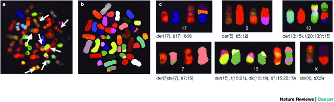
Metaphase cells from an untreated acute myelogenous leukemia (AML) patient were analyzed using spectral karyotype (SKY) analysis. a) The chromosomes were first stained with a mixture of labeled probes specific for different chromosomes. Normal chromosomes are uniform in color, whereas rearranged chromosomes show two or more colors (arrows). b) The spectral pattern of chromosomes has been classified using computer software to identify individual chromosomes. Each chromosome has its own color code. Several colors on a single chromosome indicate a rearrangement. c) Presentation of rearranged chromosomes. Each separate panel shows the spectral and classified images of the normal parent chromosome (left two chromosomes), and the spectral and classified images of the rearranged chromosome (right two chromosomes). The analysis of each chromosome type is listed below, including the chromosome that the rearrangement is derived from (der). In this cell, rearrangements are caused by translocations (t), deletions (del), or dicentric chromosomes (dic). For example, for chromosome 13 (upper right panel), the rearrangements include pieces of chromosome 15, the Y chromosome, chromosome 13, and chromosome 20, from the top to bottom. There are at least 30 separate rearrangements in this cell. Each is not necessarily associated with a cancer.


Comments
CloseComments
Please Post Your Comment